

Good horsemanship follows a solid understanding of the horse and it’s physical makeup. In order to assess the horse’s well-being you need to have good working knowledge of what you’re looking at, and what is hidden by the skin and fur.
A couple weeks ago we began our physical anatomy with Equine 101: Anatomy Part 1: Parts of the Body. Let’s get down to the bare bones and look at the skeltal system of the horse this week.
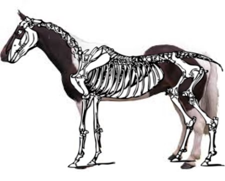
- Although imperfect, a mashup of a skeleton (pixabay.com) and a horse (wikipedia).
When I was at Equine Affaire there was a fantastic booth presenting a skeleton of an actual carriage horse (and behind it is a foal). It was fascinating to see because you could actually see some of the skeletal defects this horse presented with. If you look closely you’ll be able to see some degeneration between several of the vertebrae.
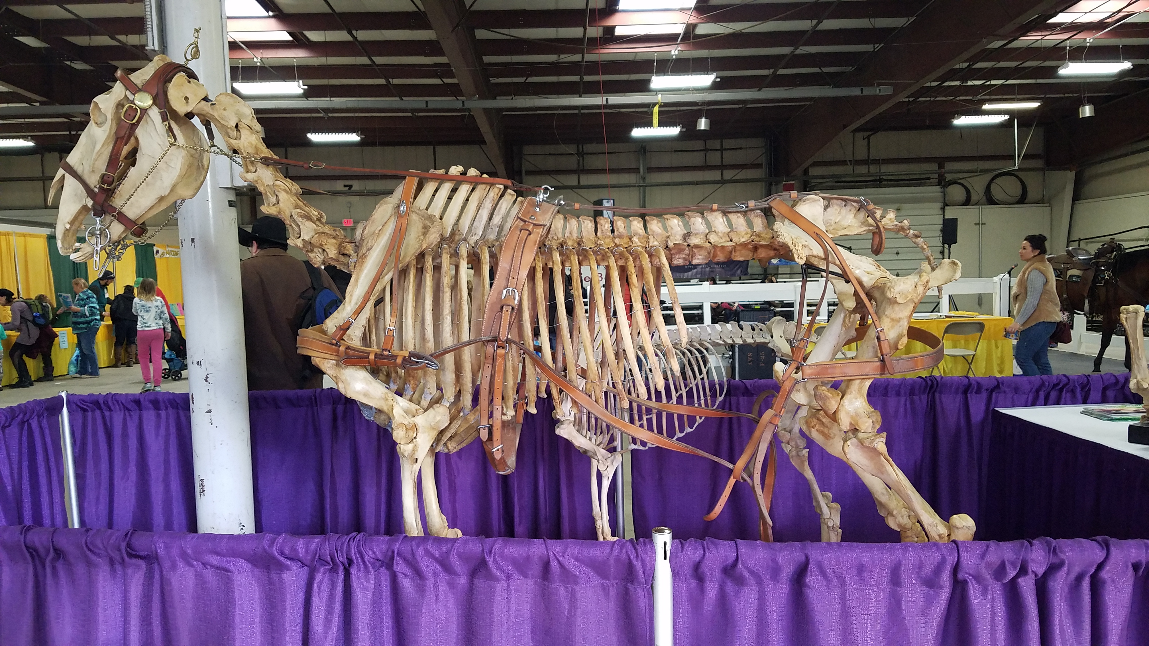
I wanted to use this photo to make a labelled diagram like I did for the body parts post, but the photo is simply too busy. Since my artistic talent is limited I am borrowing from Wikipedia this week. The most important thing I want you to take away from today is a working knowledge of how the horse moves based on it’s bone structure. When you watch a horse moving, Try to picture what the skeleton is doing.

- Photo courtesy of Wikipedia commons
The horse has about the same number of bones as humans. While we have 204, equines typically have 205. This number varies between some horses because some breeds have slightly different makeups.
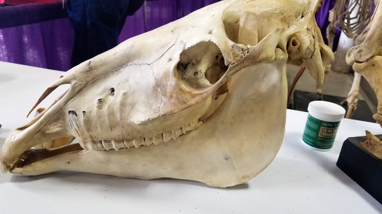
Beginning at the head we have the skull, made up of 34 bones (humans have 22). Looking at a closeup of the skull you can get a good look at the molars. Without going to dental on you the incisors are located at the front of the mouth. These are used to pick and tear at grass…and bite. Food is processed further back in the mouth by these giant molars. We’ll cover them more later.
Also seen in the skull is the large ocular orbital for the eyes and the nasal passage. You’ll notice the nasal bone gets very narrow at the end, this is where cartilage takes over. You can imagine, however, the pain it could cause by having the noseband of your bridle and halter too low on the horse’s face. Any heavy yank or jerk there could cause damage (Not that you should be yanking on your horses face all the time even with proper fitting gear).
Running back from the skull you’ll hit the vertebrae. There are 7 located in the neck and they are the cervical vertebrae. Further down from the withers to the end of the rib-cage you’ll count 18-19 thoracic vertebrae. Moving along you’ll see 5-6 lumbar vertebrae which end at the sacrum (where the pelvis begins) which consists of 5 more vertebrae that fuse together. Finally, at the tail there are 15-25 caudal vertebrae.
Fun fact: When the horse is free of tension and injuries the caudal vertebrae in the tail can actually bend up and back and touch the sacrum/pelvis area. DO NOT EVER force a horse to do this; in order to loosen the horse up I do myofascial release (topic for later). Previous injuries could also hinder a horses flexibility here.
Moving back to the front end of the horse, you’ll see the scapula. The scapula is the horse’s shoulder (they have no collar bones like humans do). When you’re fitting a saddle this is one of the most important bones (other than the thoracic vertebrae) that you need to be aware of.
The video embedded was added by Charlotte Bullard CBEST. I really love the way she has painted the skeletal anatomy to demonstrate to real time movement. Watch the scapula as it walks; as the horse moves it’s leg forward to step the top of the scapula slides backwards.
This is important to note because when you tack a horse the saddle rests over the tip of the scapula. As the horse walks the scapula needs room to slide under the flap of the saddle. If the saddle isn’t fitted properly this will cause pain and hinder the horse’s ability to extend and stretch out it’s gaits.
 Connected to the scapula is the humerus, which is mostly cutoff in the photo of the front leg. This is similar to your upper arm.
Moving down the leg you get to the ulna and radius.
Below that at the knee is the carpus, which leads to the metacarpal, or cannon bone.
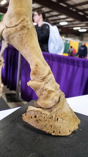
Below and behind the cannon bone is the sesamoid (fetlock bone). The longer bone under the cannon and the one below it are both pasterns. Finally at the bottom you find the coffin bone, which we talked about briefly in National Pet Obesity Awareness Day. The coffin bone sits in the hoof capsule surrounded by vessels and laminae. As the horse walks and moves the laminae absorb the pressure while distributing it through the hoof capsule.
FUN FACT: The forelimbs carry 55-60% of the horse’s body weight.
Back to the top of the horse we reach the rib-cage, horses have 18-19 ribs. Of these, 8 attach all the way around to the sternum. The rest are open to allow the body to expand for breathing.
 Most of the midsection consists of tissues and organs, so the next group in the skeletal system is the hindend. At the top, the pelvis.
The pelvis is a large area and common source of pain in many horses. The pelvis attaches to the femur, which changes direction and runs toward the front of the body.
If you run down the femur just below the breeching (thick leather strap the runs around the back end of the horse) and in front you will see a small bone. This is the patella, or knee cap. This region is the stifle (remember from the body parts page).
The tibia and fibula run back down toward the hock, which is made up of a couple tarsal bones. As in the forelimb’s metacarpal, the hindlimb has a metatarsal, which runs down to the pasterns and coffin.
I hope this helps you look at your horse in a different way and provides you with the beginning knowledge of what goes on beneath the skin. Although it seems lengthy above this just scratches the surface. In time I plan to look at specific locations so we can see what goes on at each area.


Pictures above courtesy of Wikipedia.
As always I want to leave you with some extra reading material. These resources have helped me in my learning as I’m sure they’ll help you.
HorseChannel.com – 7 Facts About Your Horse’s Skeleton


Leave a comment